

Guided Implant Planning (CBCT-Based)

The dentist's intraoral scan registered to the CBCT, implants positioned to serve the final restoration, and the plan handed straight to the surgical guide.

A CBCT tells you where the bone is. It does not tell you where the implant belongs — that is a restorative question, and answering it in software before surgery is the whole point of CBCT-based implant planning.

Jahn De Khudikine, Founder · Universal Dental Lab
Jahn De Khudikine Founder · Universal Dental Lab

Why send it here

Plan where the restoration wants the implant — before anything is cut.

A practice's schedule lives or dies by the lab — I've watched one late case push back a whole day of chair time.

So I still know the cases moving through this lab, and I answer for how each one is made. Send a case here and it doesn't drop into a queue — it lands with me and one technician who carries it start to finish.

Start a case
The restoration leads
Implants positioned to serve the final prosthesis Screw access and emergence decided in software, where changing them is free
You approve first
Nothing downstream is built until you sign off The plan you review is the file the guide is built from
Your technician
One planner holds your case Reach them directly — no call-center queue
Free scanner
3Shape TRIOS 6 for partners Go fully digital at no cost
Universal Dental Lab Planning records

What to send with the case

  • CBCT scan (DICOM)
  • Intraoral scan, both arches (STL)
  • Bite registration
  • Implant system and platform
  • Planned restoration type
  • Existing prosthesis or wax-up, if relevant

The closer in time the CBCT and the scan were taken, the cleaner the alignment.

How We Work With You

Plan Where the Restoration Wants the Implant

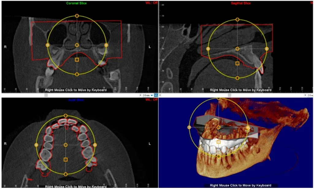
We import your DICOM, register the intraoral scan to it, and separate bone, teeth and soft tissue. Then we set the final restoration and place each implant to serve it: angulation that keeps screw access off the incisal edge, depth that gives a workable emergence profile, and clearance to the structures you cannot afford to hit.

Vital structures first
The inferior alveolar canal, the sinus floor, adjacent roots and the buccal plate are identified and given margin before any implant is positioned. When a site is tight, we show you the constraint rather than quietly shaving the safety margin to make a position work.
The restorative question
Restorative space is the most common surprise — a position that works in bone but leaves nowhere for the prosthesis. Angulation is next: a site that allows a screw-retained crown at one inclination and forces cement at another. We present both options and the trade-off instead of picking one silently.
Plan to guide
On approval, the positions you signed off on export straight into surgical guide design — the guide is built from the same file you reviewed, not re-planned from a screenshot.
CBCT segmentation on screen with the inferior alveolar canal traced and implant positions placed to the restoration

The Read

What We Map Before an Implant Is Positioned

We import your DICOM, register the intraoral scan to it, and separate bone, teeth and soft tissue. Then we set the final restoration and place each implant to serve it: angulation that keeps screw access off the incisal edge, depth that gives a workable emergence profile, and clearance to the structures you cannot afford to hit.

  • Vital structures: The inferior alveolar canal, sinus floor, adjacent roots and buccal plate are identified and given margin before any implant is positioned.

  • Restorative space: The most common surprise — a position that works in bone but leaves nowhere for the prosthesis. We show the constraint instead of shaving the margin.
Learn more
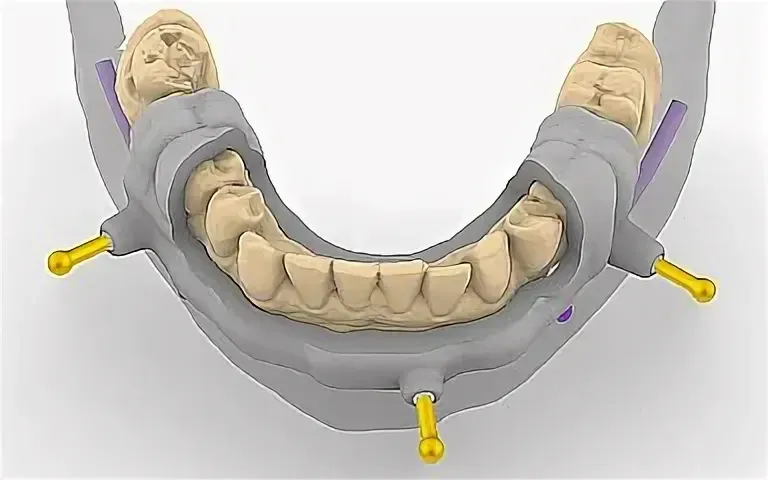
Surgical guide design derived from the approved implant plan, sleeve positions in place on the model

From Plan to Guide

The Positions You Approve Are the Ones We Build

Once you approve the implant positions, they export straight into surgical guide design — the guide is generated from the same plan you reviewed, not re-created from a screenshot. Sleeve position and drill offset inherit the angulation and depth you signed off on.

For full-arch cases that need vertical reduction, the same CBCT drives the bone reduction guide, so the planning, the reduction and the placement stay on one coordinate system.

  • One file: The guide is generated from the same plan you reviewed — nothing is re-interpreted between planning and fabrication.

  • One coordinate system: For full-arch work the same CBCT drives the reduction guide, so planning, reduction and placement stay aligned.

Learn more

Full-arch and complex cases

Full-arch and hybrid cases carry a heavier planning load and less room to improvise. They run through the same segmentation and prosthetically driven workflow with added prosthetic staging — see full arch case planning for how All-on-X work is handled, and bone reduction guides for the reduction step planned from the same CBCT.

The Process

How a CBCT Case Is Planned

From two datasets to an approved set of positions. Nothing is fabricated until you sign off.

  1. Send the CBCT and the scan

    Practice

    A CBCT in DICOM and an intraoral scan of both arches in STL, taken close enough together that the anatomy still agrees. Add the implant system, the planned restoration, and any prosthesis the plan should respect.

  2. We segment and align

    Lab

    We import the DICOM, register the intraoral scan to the CBCT, and separate bone, teeth, and soft tissue. If the two datasets do not line up — motion artifact, scatter from restorations, a scan taken months apart — you hear about it before we plan.

  3. Prosthetically driven positioning

    Lab

    We set the final restoration first, then position each implant to serve it: angulation for screw access, depth for emergence, and safe margins to the sinus floor and inferior alveolar canal. Bone constrains the plan; it does not dictate it.

  4. You review and approve

    Practice

    You get the plan with proposed positions, the restorative rationale, and the trade-offs where a site allows more than one answer. Nothing downstream is built until you sign off.

  5. Plan becomes a guide

    Lab

    On approval the approved positions flow straight into surgical guide design, so the guide is built from the same file you reviewed — not re-planned from scratch.

What CBCT-based planning actually buys you

  • Vital structures mapped first

    The inferior alveolar canal, sinus floor, and adjacent roots are identified and given margin before an implant is ever positioned, so the plan respects them by design rather than by luck.

  • The restoration leads

    Implants are placed to serve the final prosthesis — screw access, angulation, and emergence are decided in software, where changing them is free.

  • One file, plan to guide

    The positions you approve are the positions the surgical guide is built from. Nothing is re-interpreted between planning and fabrication.

Start a case

Send one case. Keep the scanner.

Open a partner account and your first case moves with a shade record and our remake guarantee — if a unit does not seat, we redo it at no charge.

  • Free 3Shape TRIOS 6 scanner for active partners
  • Under-2% remake rate — remakes at no charge
  • One named technician you reach directly — no call center

Prefer to talk? +1 747-268-0808

No call center — you hear back from the lab, not a bot.

Planning FAQ

Guided Implant Planning — Questions Dentists Ask

Often yes if the dentition and soft tissue have not changed materially. The closer in time and the cleaner both datasets, the more reliable the registration. If the patient lost a tooth, had a provisional remake, or the scan captures different jaw position, tell us — we may ask for an updated scan before implant positions are finalized.

System, platform, and the intended restoration — single crown, bridge span, or full-arch hybrid. Planned drill kit or guided protocol matters when the plan will become a guide. Without platform clarity we cannot validate components or screw-access angles in the virtual plan.

How to send a file

Heavy scatter from existing metal restorations can degrade segmentation; motion blur and limited field of view do the same. We will tell you if the CBCT is workable or if a new scan protocol — smaller arch, different settings, or scan after provisional metal removal — is needed before you schedule surgery.

Proposed implant positions tied to the restorative plan, notes on angulation and access, and flags on thin bone or inadequate prosthetic space while changes are still cheap. Approved plans feed surgical guide design and custom abutment CAD — this service is the planning layer, not the printed guide itself.

Surgical guide design

No — this page is prosthetically driven planning on your CBCT and scan. The guide product pages cover fabricated guides and stacked full-arch sequences. Plan here first; once positions are approved, guide design turns the plan into sleeves, stops, and printable geometry.

Surgical implant guides

Partner Offer

A Free Intraoral Scanner for Partner Practices

Partner with Universal Dental Lab and we place a 3Shape TRIOS 6 Wireless in your operatory — free for the length of our partnership. Scan the prep, send it in seconds, and skip impressions entirely.